nikon small world announces 2022 photomicrography winners
Nikon Small World has recently announced the top 20 winners of its 2022 Photomicrography Competition, sharing a new yearly series of dazzling images, each spotlighting the artistry of the microscopic realm. With approximately 1,300 entries submitted from 72 countries, the 2022 winning round also marks the competition’s 47th anniversary, first launched in 1975.
‘[It] first began in 1975 as a means to recognize and applaud the efforts of those involved with photography through the light microscope. Since then, Small World has become a leading showcase for photomicrographers from the widest array of scientific disciplines,’ writes Nikon Small World.
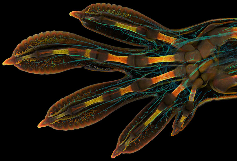
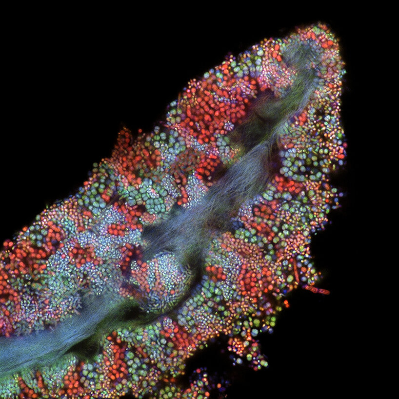
1st place: embryonic hand of a Madagascar giant day gecko (Phelsuma grandis) | 63X (magnification)
image by Grigorii Timin & Dr. Michel Milinkovitch | all courtesy of Nikon Small World
a yearly celebration of a world invisible to the naked eye
From fluorescent depictions of tissue cells and blood vessels to magnified shots of insects and microscopic organisms, the Nikon Small World 2022 Photomicrography Competition gives viewers a yearly glimpse into a fascinating and incredibly rich world that is invisible to the naked eye. Participants include university professors, practicing scientists, nature enthusiasts / photographers, and PhD students. You can view more winning images from previous years on the Nikon Small World website.
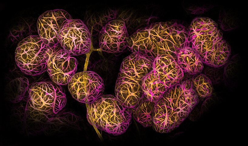
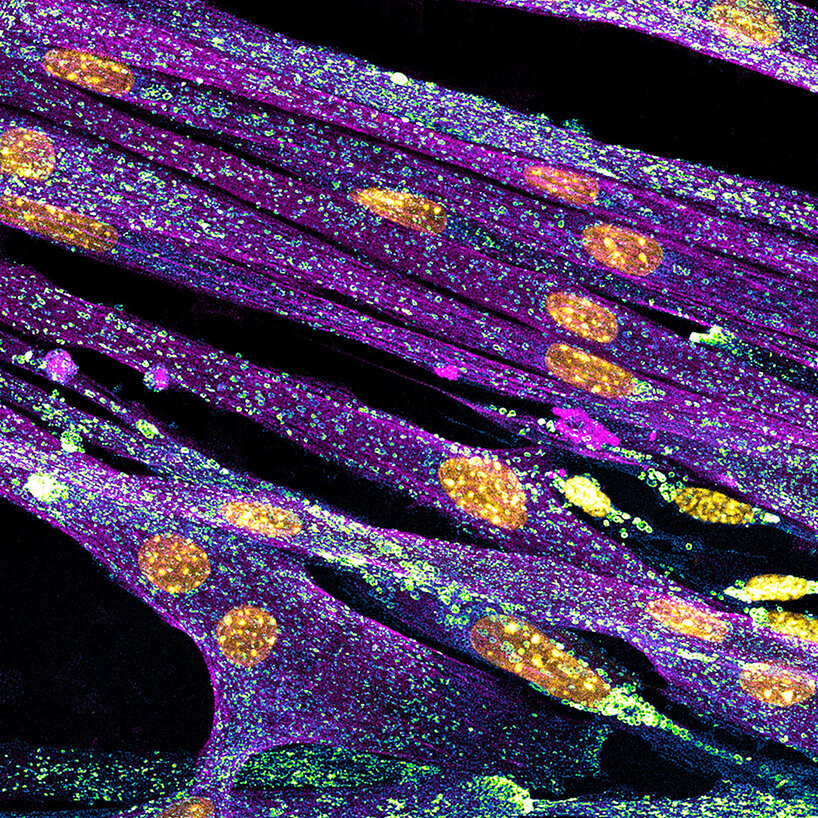
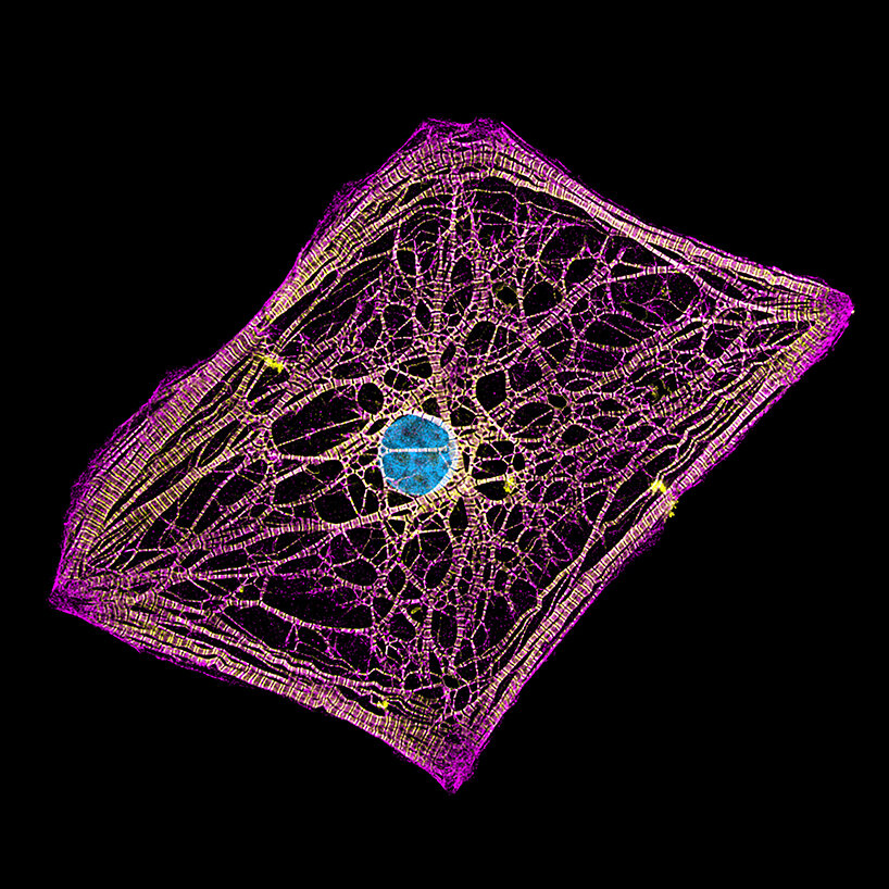
2nd place: breast tissue with contractile myoepithelial cells wrapped around milk-producing alveoli | 40X
image by Caleb Dawson
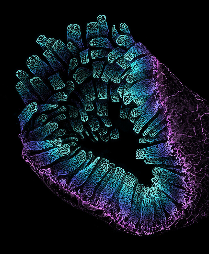
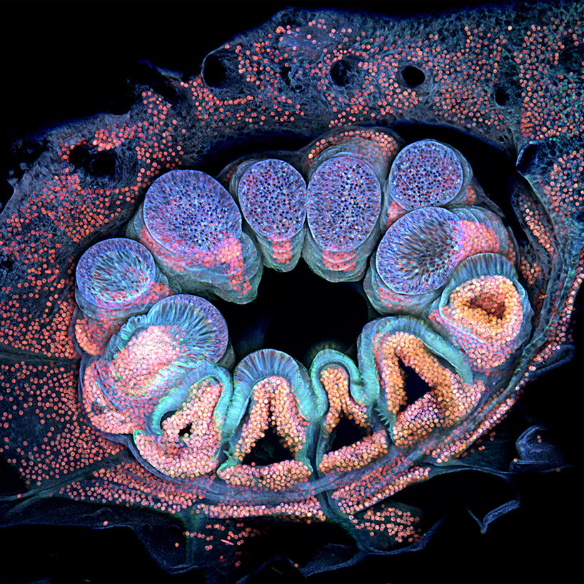
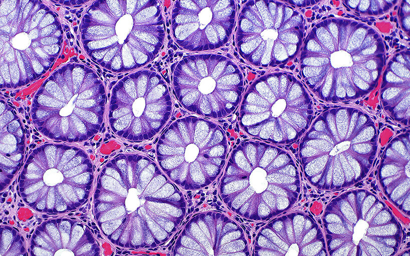
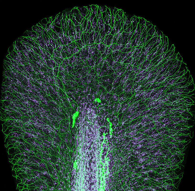
3rd place: blood vessel networks in the intestine of an adult mouse | 10X
image by Satu Paavonsalo & Dr. Sinem Karaman
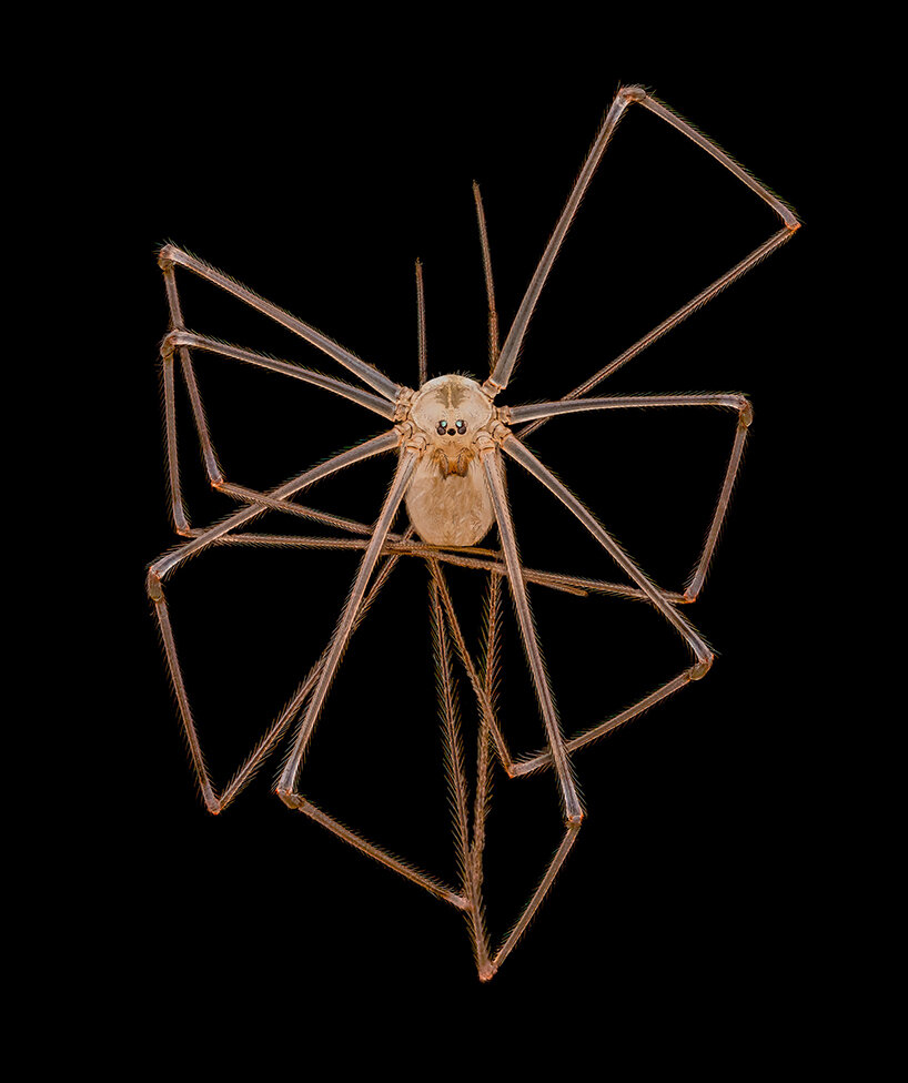
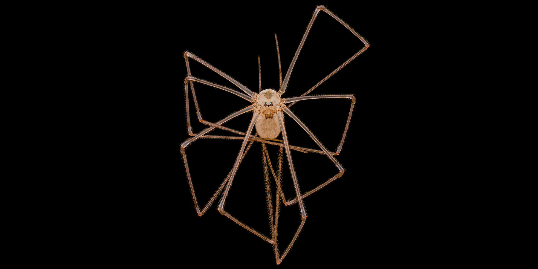
4th place: long-bodied cellar/daddy long-legs spider (Pholcus phalangioides) | 3X
image by Dr. Andrew Posselt
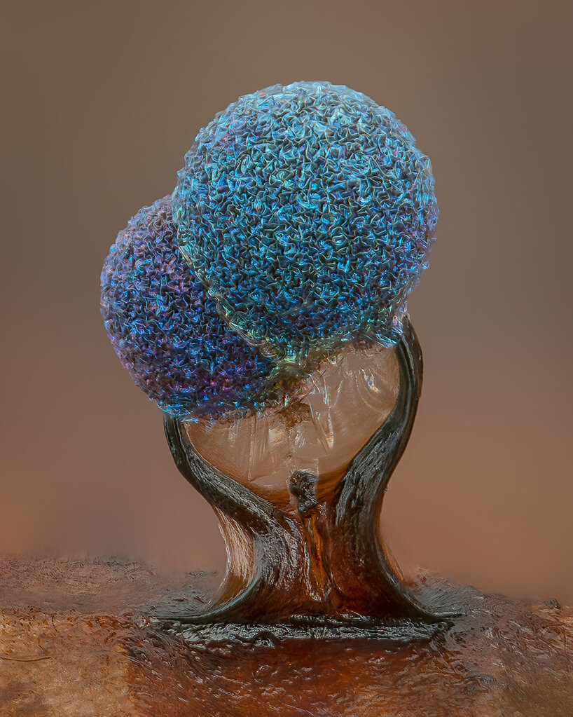
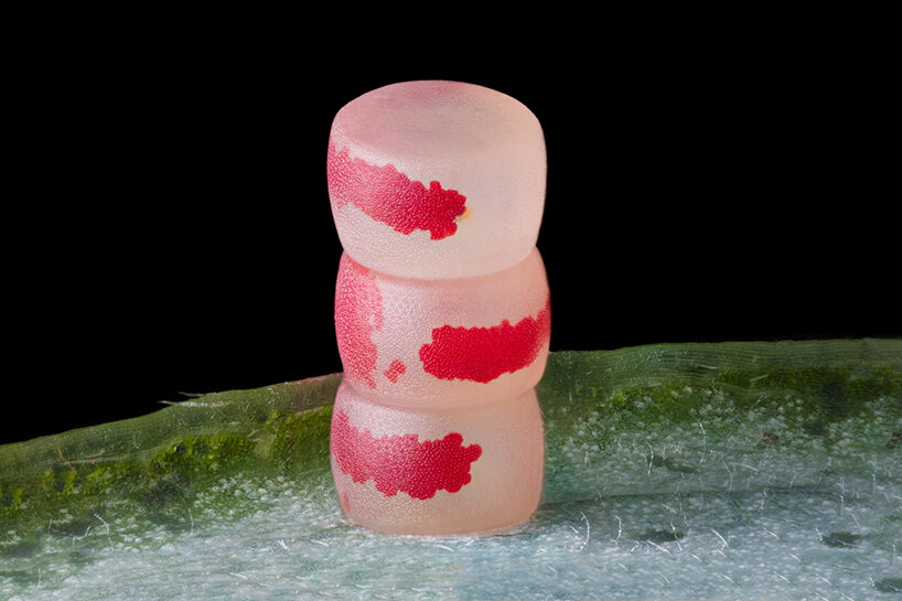
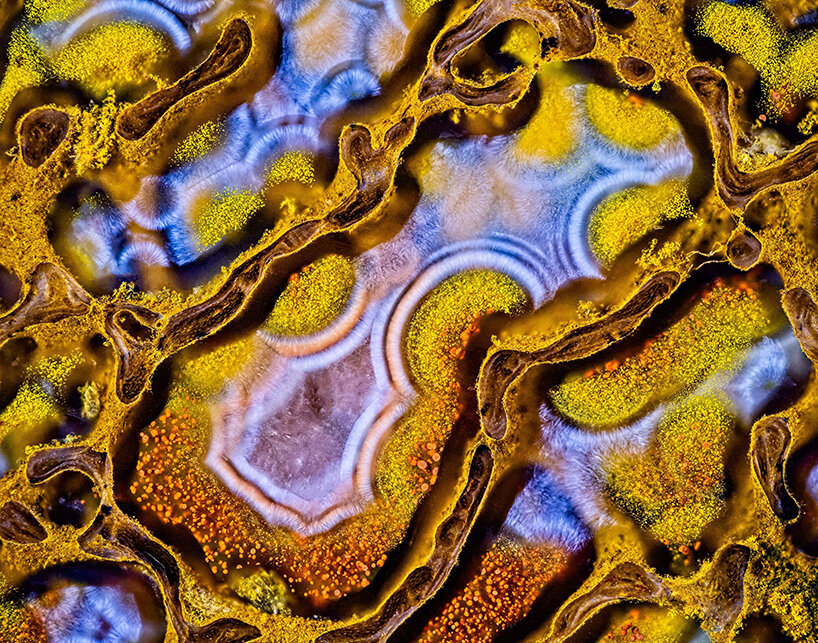
5th place: slime mold (Lamproderma) | 10X
image by Alison Pollack

6th place: unburned carbon particles released during breakdown of hydrocarbon candle wax chain | 2.5X
image by Ole Bielfeldt
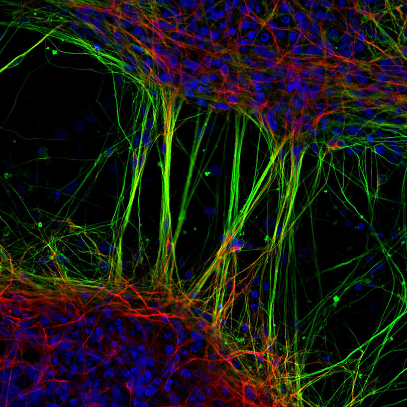
7th place: human neurons derived from neural stem cells (NSCs) | 20X
image by Dr. Jianqun Gao & Prof. Glenda Halliday
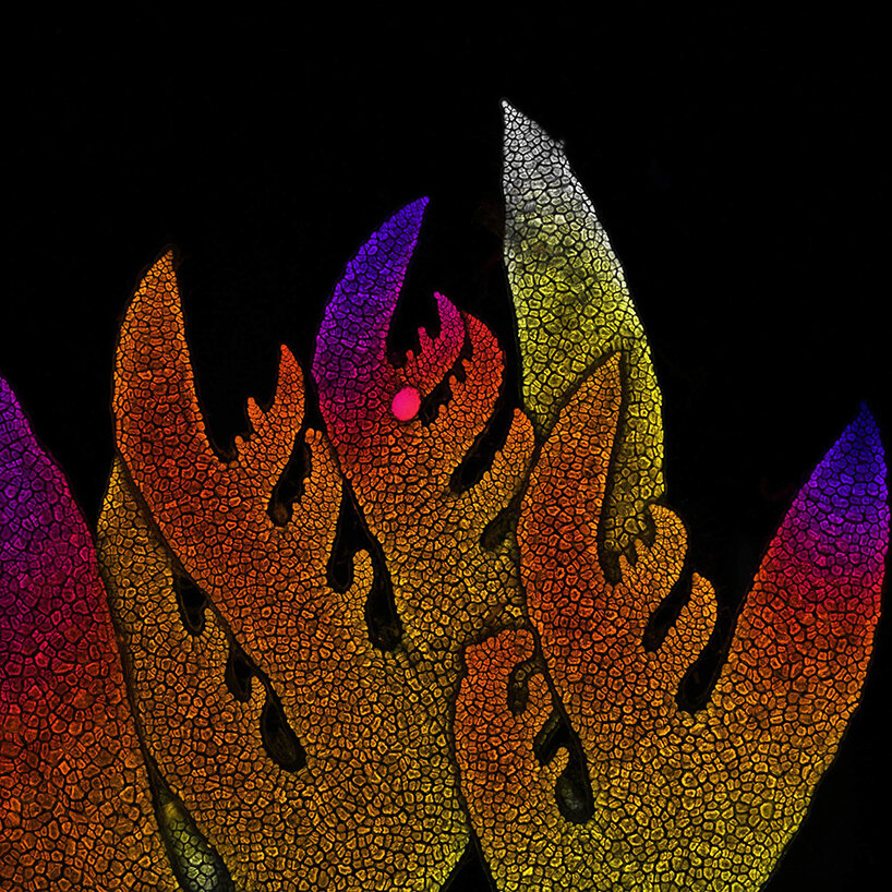
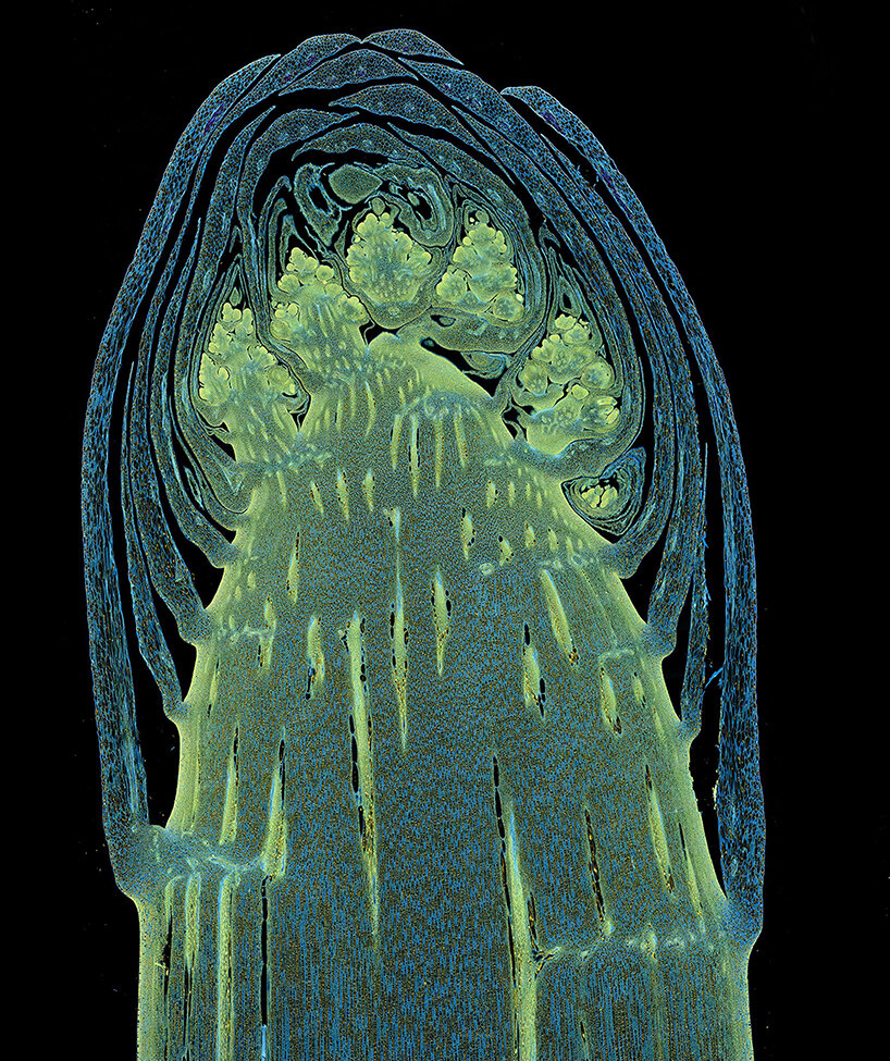
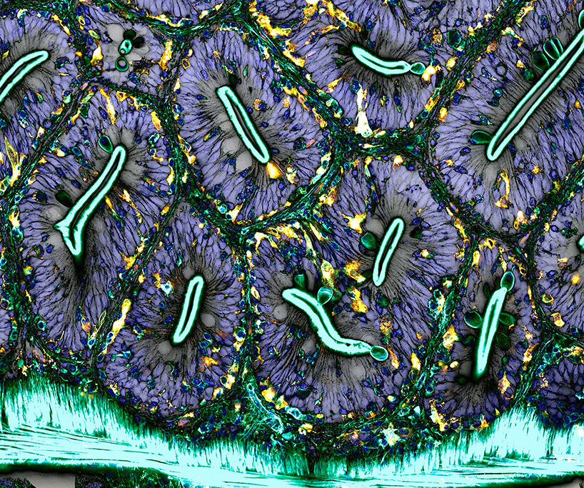
8th place: growing tip of a red algae | 10X
image by Dr. Nathanaël Prunet
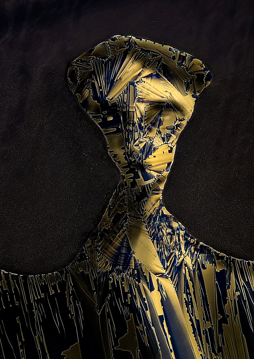
9th place: Liquid crystal mixture (smectic Felix 015) | 40X
image by Marek Sutkowski
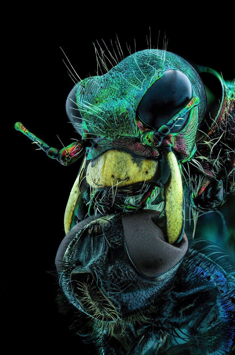
10th place: A fly under the chin of a tiger beetle | 3.7X
image by Murat Öztürk
project info:
name: 2022 Photomicrography Competition
organized by: Nikon Small World
nikon (11)
 Apr 02, 2020
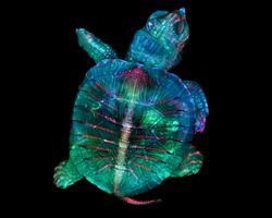 Oct 22, 2019
 Jul 11, 2018
 Aug 17, 2016
 Aug 02, 2016
PRODUCT LIBRARY
 Jul 15, 2024
 Jul 15, 2024
 Jul 12, 2024