the winning photographs in nikon‘s small world photography competition include a disco-hued octopus embryo and a closeup of a housefly‘s eye. the competition, which highlights the best
microscope images taken each year, attracted more than 2,000 entries from nearly 100 countries.

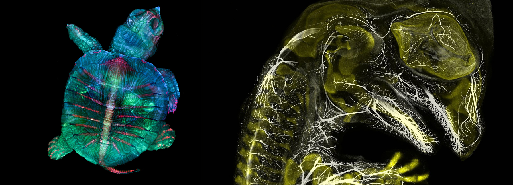
2nd place, depth-color coded projections of three stentors (single-cell freshwater protozoans), by dr. igor siwanowicz / cover image: 1st place, fluorescent turtle embryo by teresa zgoda and teresa kugler
images courtesy of nikon small world
the winner of the 2019 edition was microscopy technician teresa zgoda and recent university graduate teresa kugler who captured a developing turtle embryo, stained to reveal different tissues and shown at fives time magnification. second place was awarded to nikon small world veteran dr. igor siwanowicz for his composite image of three single-cell freshwater protozoans, sometimes called ‘trumpet animalcules.’ he used confocal microscopy to capture the detail of the cilia, tiny hairs used by the animals for feeding and locomotion.

3rd place, alligator embryo developing nerves and skeleton, by daniel smith paredes and dr. bhart-anjan s. bhullar
third place was awarded to mr. daniel smith paredes, who placed for his image of a developing american alligator embryo. he snapped this photo at around 20 days of development using immunofluorescence and is studying the development and evolution of vertebrate anatomy. other entries featured in the top 20 included a cross-section of a tulip bud, tiny single-cell freshwater protozoans and a focus-stacked image of a small spider.

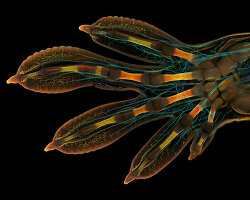
4th place, male mosquito, by jan rosenboom
the nikon international small world photography competition first began in 1975 as a means to recognize and applaud the efforts of those involved with photography through the light microscope. since then, small world has become a leading showcase for photomicrographers from the widest array of scientific disciplines.

5th place, snowflake, by caleb foster
‘microscopy lets us zoom in on the smallest organisms and building blocks that comprise our world – giving us a profound appreciation for the small things in life that far too often go unnoticed,’ said kugler, ‘it allows me to do science with a purpose.’

6th place, small white hair spider, by javier rupérez
‘we are inspired by the beautiful images we see through the microscope,’ added zgoda, ‘it’s humbling and deeply fulfilling to be able to share that science with other people.’

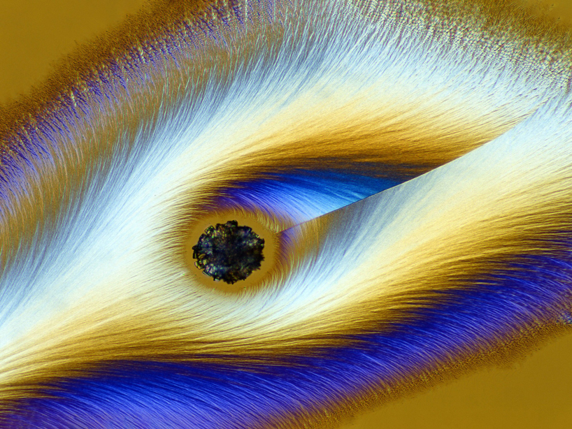
7th place, chinese red carnation stamen, by dr. guillermo lópez lópez
‘our goal has always been to show the world how art and science intersect,’ says eric flem, communications manager, nikon instruments. ‘as new imaging and microscopy techniques develop over the years, our winners showcase these technology advances more and more creatively.’

8th place, frozen water droplet, by garzon christian

9th place, tulip bud cross section, by andrei savitsky

10th place, BPAE cells in telophase stage of mitosis, by jason m. kirk
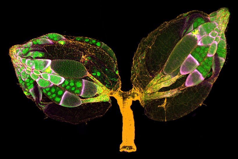
11th place, a pair of ovaries from an adult drosophila female stained for f-actin (yellow) and nuclei (green); follicle cells are marked by GFP (magenta), by dr. yujun chen and dr. jocelyn mcdonald
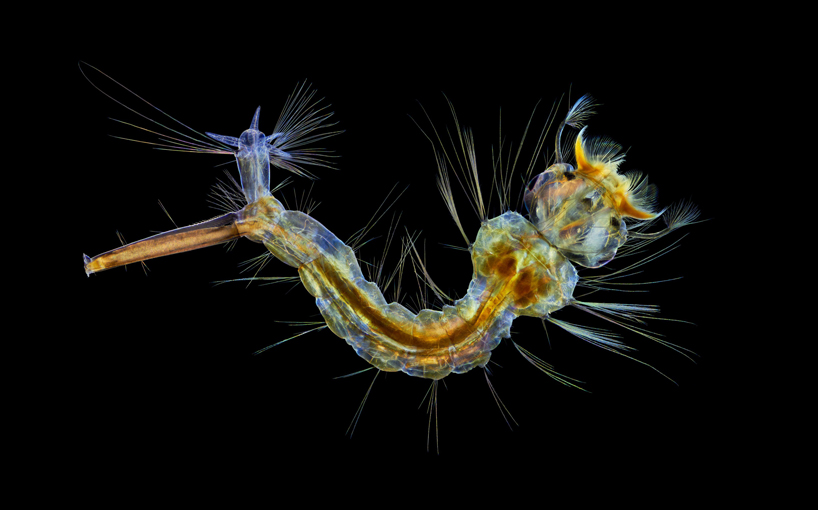
12th place, mosquito larva, by anne algar

13th place, cuprite (mineral composed of copper oxide), dr. emilio carabajal márquez
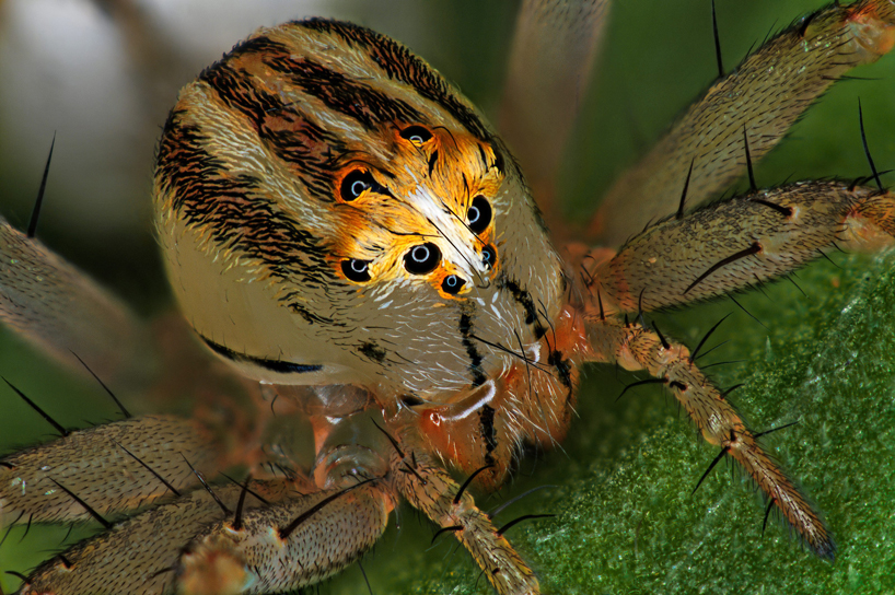
14th place, female oxyopes dumonti (lynx) spider, by antoine franck
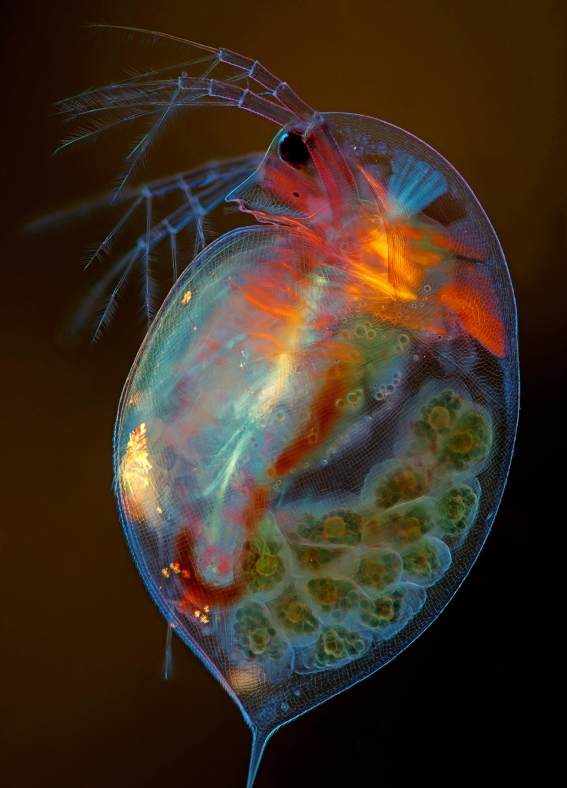
15th place, pregnant daphnia magna (small planktonic crustacean), by marek miś
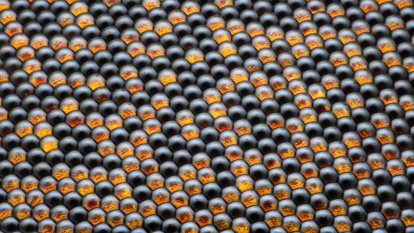
16th place, housefly compound eye pattern, by dr. razvan cornel constantin
 17th place, vitamin c, by karl deckart
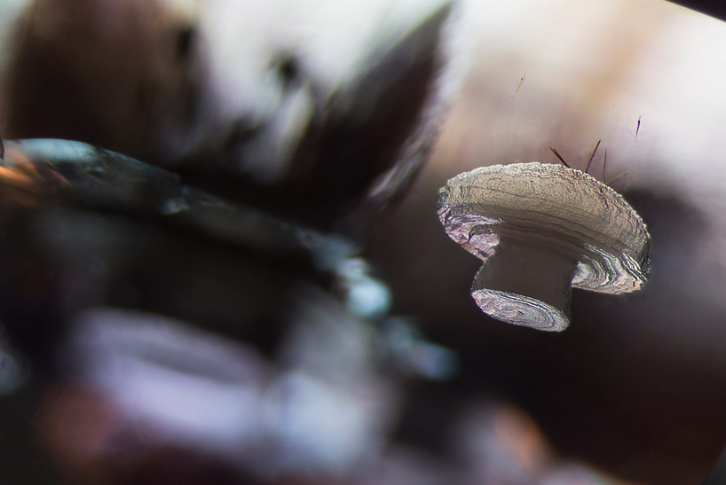
18th place, cristobalite crystal suspended in its quartz mineral host, by e. billie hughes
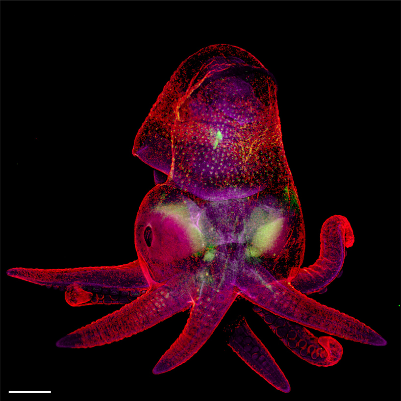
19th place, octopus bimaculoides embryo, by martyna lukoseviciute and dr. carrie albertin
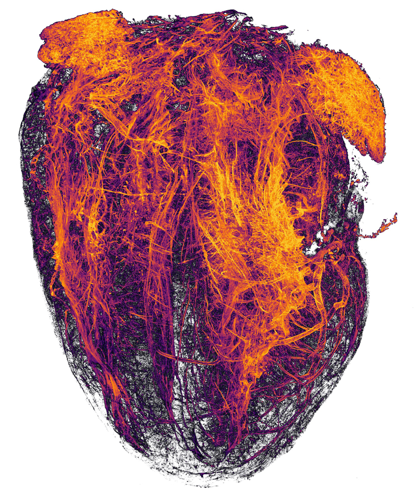
20th place, blood vessels of a murine (mouse) heart following myocardial infarction (heart attack), by simon merz, lea bornemann and sebastian korste
project info:
company: nikon
competition: nikon small world photography
type: photography
insects (36)
 Jun 25, 2024
 Apr 02, 2024
 Aug 10, 2023
 Nov 10, 2022
 Nov 02, 2022
nikon (11)
 Oct 24, 2022
 Apr 02, 2020
 Jul 11, 2018
 Aug 17, 2016
 Aug 02, 2016
photography (389)
 Jul 18, 2024
 Jul 16, 2024
 Jul 15, 2024
 Jul 08, 2024
 Jul 02, 2024
PRODUCT LIBRARY
 Jul 15, 2024
 Jul 15, 2024
 Jul 12, 2024