Google and harvard researchers generate human brain images
Researchers from Google and Harvard University have recently released a new set of detailed images of the human brain and its neural networks using AI and 3D mapping. The research is ten years in the making with Google Research’s Connectomics research team, in collaboration with Jeff Lichtman of Harvard University and others. A large dataset was gathered by the Harvard University researchers using a donated brain sample that was removed during surgery on a woman with epilepsy. Jeff Lichtman and his team used a multibeam scanning electron microscope to gather high-resolution images of more than 5,000 slices of tissue.
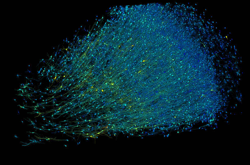
This process alone took 326 days. Google’s research team and its AI and 3D mapping stepped in here. They pieced the image data together and generated them into a 3D structure detailing each cell. By combining brain imaging with AI-based image processing and analysis, Google and Harvard University researchers managed to reconstruct nearly every cell and all of its connections within a small volume of the donated human brain tissue, which was about half the size of a grain of rice, and produced images that could help researchers understand neurological disorders and learn more about how the brain works.
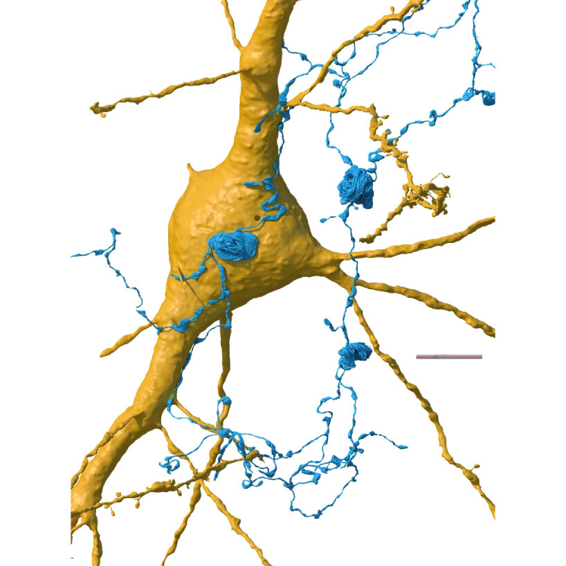
Axon whorls | images courtesy of Google Research & Lichtman Lab (Harvard University) | renders by Daniel Berger
Neural networks come to detailed life using AI and 3D mapping
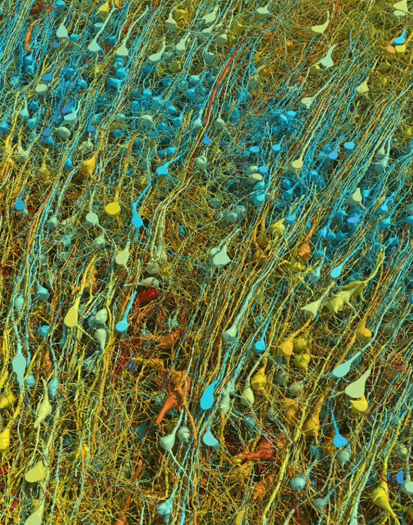
The neural network image made using AI and 3D mapping may be the largest dataset ever made of human brain structure. Google’s research team says that illustrating this small part of the donated human brain already takes more than a million gigabytes of data, or 1.4 petabytes. The sample used for the high-resolution photo came from a part of the cortex called the anterior temporal lobe, which has six layers. Google and Harvard University researchers added colors to the neurons according to their size and type to make their layers visible and easily identifiable for studies.
The Harvard University and Google researchers used a tiny tissue sample, about the size of one cubic millimeter, and discovered several numbers of cells and connections. It contained around 50,000 cells, which are the basic building blocks of the nervous system, and there were about 150 million synapses, or the points where signals are transmitted between neurons, or nerve cells, in the brain. Some pairs of neurons in the sample were found to be connected to each other very strongly, and in some cases, they were connected by as many as 50 synapses. So far, researchers are unsure why these connections are so strong and are looking into them.
one cubic millimeter tissue sample contained about 50,000 cells and about 150 million synapses
Researchers discovered in the human brain
There are several surprises that were in store for Google and Harvard University researchers, as indicated in their published study. The use of AI and 3D mapping allowed them to see the occurrence of ‘axon whorls’. For context, axons are like long wires that extend from neurons and carry signals away from the cell body, playing an important role in transmitting information throughout the nervous system. In the tissue sample, the researchers found some of them looped or coiled instead of extended. These structures were not common and were even found in a few instances within the sample.
In some cases, these axon whorls were observed sitting on the surface of another type of cell. Researchers are not sure yet what the exact function or purpose of these axon whorls is, and it’s still unclear why they form or what role they play in the nervous system. The researchers also found green axons signaling to the neuron to send ‘excitatory signals’ and a series of blue axons signaling to the neuron not to send excitatory signals. In a nutshell, the neuron receives signals from many axons, some telling it to accept and others telling it not to.
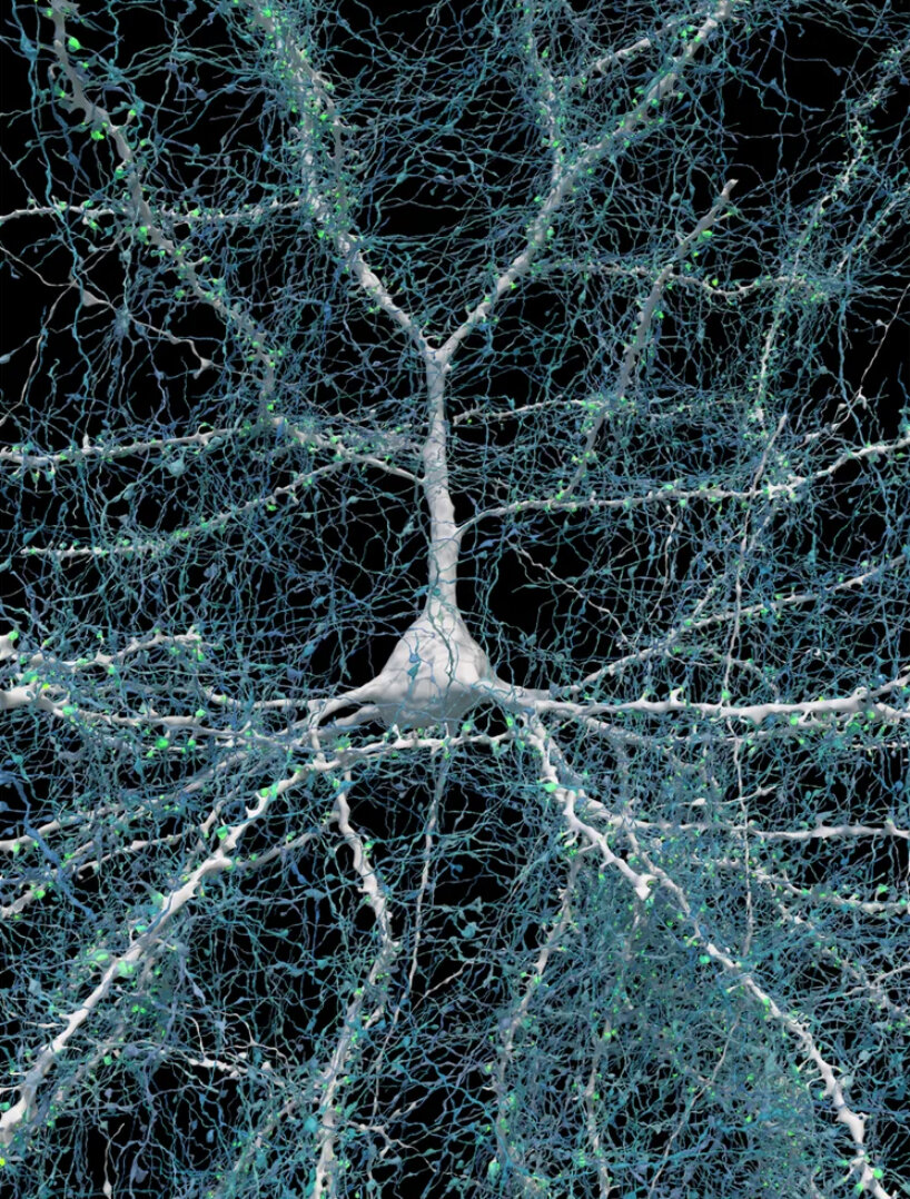
neurons in the brain are intensely connected
Multiply this process by the billions of neurons in the brain, and this shows the immense amount of communication and activity happening in the brain all the time. Since the release of the detailed human brain image using AI and 3D mapping, the researchers are now hoping to make use of the data for additional discoveries.
They’re looking into the brain’s connections next and aim to understand how memories are formed, what leads to neurological disorders and diseases like autism and Alzheimer’s, and more. This is already in the works by investigating the hippocampal formation of a mouse. Google and the other researchers have already partnered with the team at the Institute of Science and Technology Austria for the research.
Google and Harvard researchers show image of inhibitory neurons in human brain using AI and 3D mapping
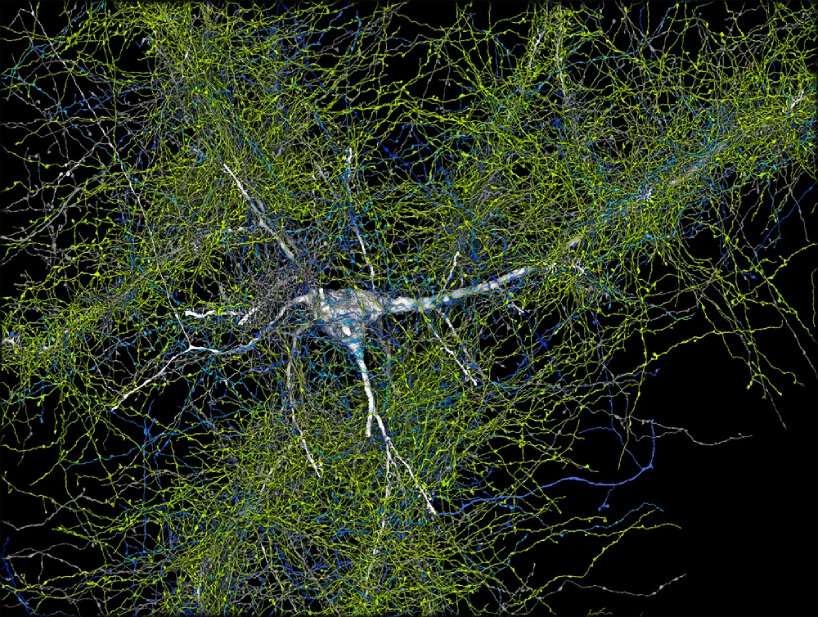
the neuron receives signals from many axons, some telling it to accept and others telling it not to

Google and Harvard researchers combine human brain imaging with AI processing and analysis and 3D mapping
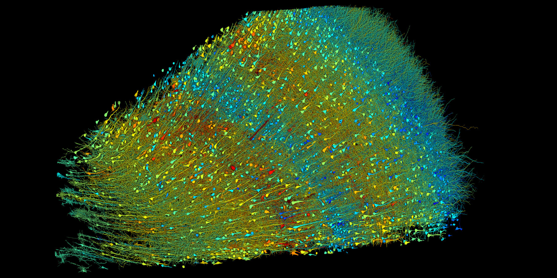
neural networks made using AI and 3D mapping may be the largest dataset ever made of human brain structure
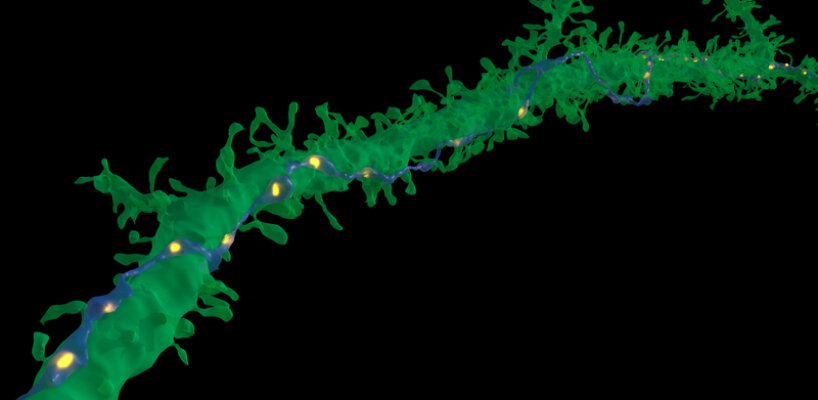
in very rare cases, a single axon (blue) made repeated synaptic connections (yellow) with a target neuron (green)
the connectome for the ‘hemibrain’ of the fruit fly
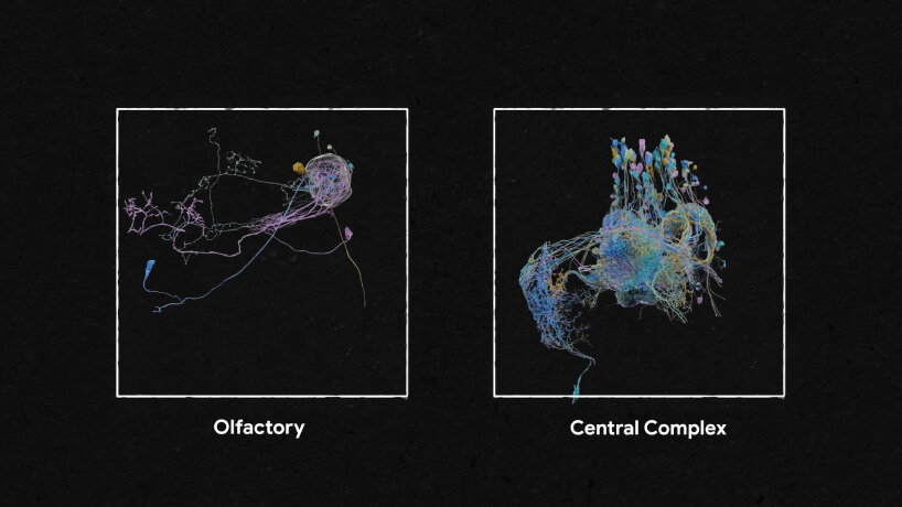
olfactory and central complex neural networks
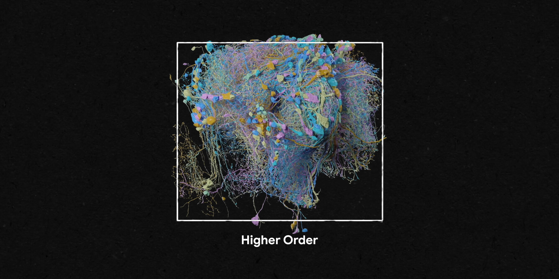
‘higher order’ image generated using AI and 3D mapping by Google and Harvard reseachers
artificial intelligence (378)
 Jul 22, 2024
 Jul 19, 2024
 Jul 16, 2024
 Jul 11, 2024
google (133)
Jul 11, 2024 May 27, 2024
 Apr 16, 2024
 Jan 06, 2024
 Nov 10, 2023
PRODUCT LIBRARY
 Jul 22, 2024
 Apr 22, 2024
 Mar 21, 2024
 Mar 13, 2024